GUINIER DIFFRACTOMETER
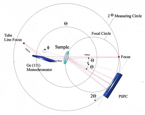
Beampath in monochromatic transmission x-ray powder diffractometry (Guinier-focussing) with PSPC.
General Description
The transmission diffractometer with focussing beampath after Guinier works with purely
monochromatic Cu-Kα1-radiation. A focussing Ge(111) monochromator (Huber Type 616-2) with
a strongly asymmetric cut of the crystal provides a beam cross section of 5 mm (beam hight 12 mm)
at the sample integrating over a suffiently large number of powder particles to meet the necessary
crystallite statistics for good “powder“ data. In addition samples can be rotated and a
position-sensitive detector collects all diffracted beams into a 2θ range of 10°. Altogether the
reciprocal space is spiralled with a broad band, so that extremely high data quality from both
resolution and crystallite statistics are produced in a minimum of time.
The technique is therefore not only useful for fast and well separable phase identifications but
also for advanced crystallographic evaluations including indexing, line profile analysis and structural
refinements using the “Rietveld“ full-pattern analysis. The 2θ range was especially cultivated in
the front angle region, so that d-values of nearly 10 nm (2θ = 1°) can be measured precisely. During
the past nearly 20 years the Guinier-Diffractometer at LabXA
was used very intensively with more than 10000 powder samples.
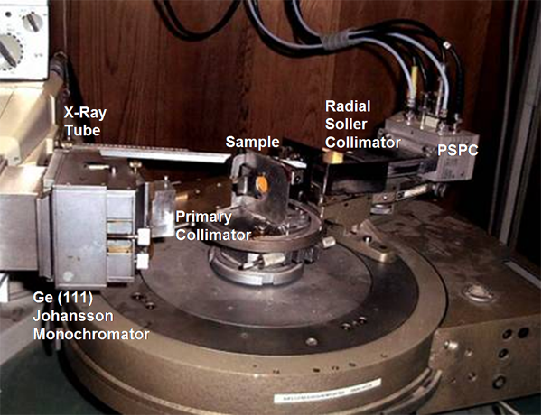
Guinier-focussing transmission diffractometer with PSPC.
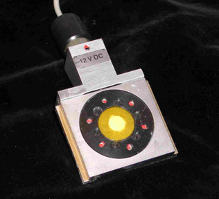
Sample rotation stage allows to maximize the crystallite statistics
Applications
The Guinier diffractometer of LabXA is designed for low-cost
routine phase analysis of crystalline powders. In the early years of LabXA it was used to analyse
thousands of kidney stone fragments for the University Clinics Munich during the development of the
shock wave disintegration of these stones (ESWL, Example 1). The high speed and data quality have
made it the “work horse“ for the analysis of powders at LabXA even for sophisticated crystallographic
evaluations like indexing and Rietveld structural analysis (see examples 2,4,5).
The accurate measurements of low diffraction angles has been a problem in powder diffraction due to
intersections with the primary beam. Special efforts were put into the beam path to allow perfect
measurements of d-values up to 10nm as they are observed for layered organic structures (stearates),
pharma products and clay minerals.
| Example 1: | Rapid high quality data collection for routine phase analyses (medical application: identification of a kidney stone). |
| Example 2: | Rapid Scan (30° / min) with PSPC. With sample rotation and continuous PSPC scan a high solid angle (see red line) of the reciprocal space (crystallite statistics) is covered. |
| Example 3: | Phase Separation in line-rich patterns |
| Example 4: | Detection at very small diffraction angles necessary for the registration of characteristic basal lines in Lead Stearate. |
| Example 5: | Excellence of data collected by our Guinier system: Rietveld refinement of triclinic AlVO4 (sample: H. Knözinger, LMU Munich, data: LabXA, Rietveld refinement: A. Kern, Bruker AXS). |
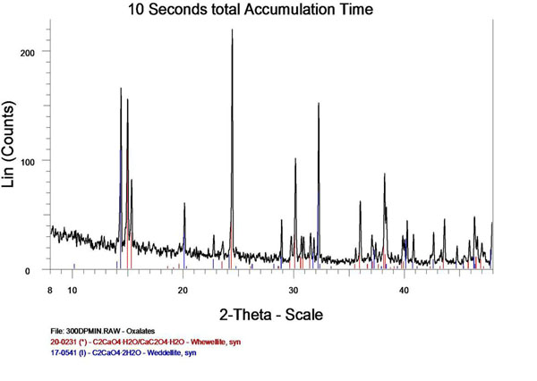
Example 1: Rapid high quality data collection for routine phase analyses (medical application: identification of kidney stone).
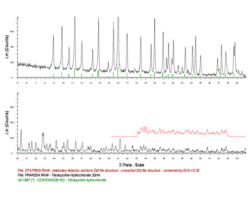
Example 2: Rapid Scan (30° / min) of Tetracycline Hydrochloride (Aldrich). The material is used by PANalytical as demo-sample to show the performance of their X’Cellerator system, however, with nearly 10 times slower scan rate. The higher speed at LabXA is a consequence of a wider angular range (10° at .01° resolution = 1000 channels, red insert) simultaneously observed by the PSPC.
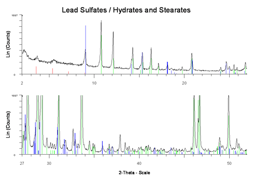
Example 3: Phase-Identification in line-rich patterns require high resolution, monochromatic profiles, high accuracy, low background and a sufficient angular range (here a mixture of triclinic. monoclinic and hexagonal lead compound used as fillers in PCV polymers)
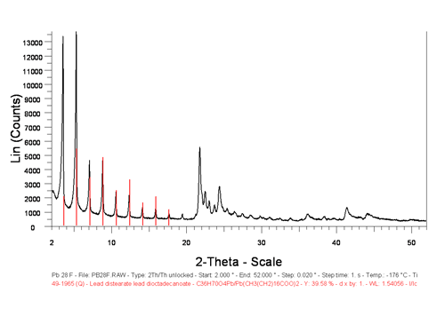
Example 4: Detection at very small diffraction angles necessary for the registration of characteristic basal lines in Lead Stearate.
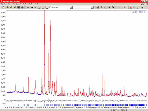
Example 5: Excellence of data collected by our Guinier system: Rietveld refinement of triclinic AlVO4 (sample: H. Knözinger, LMU Munich, data: LabXA, Rietveld refinement: A. Kern, Bruker AXS).